BI for CELLULAR STUDIES
3.5.13.
The initial idea of culturing cells within a channel of an FI system, (Ruzicka 1992) with the aim of exploring cellular reactions to impulses of stimulant was naïve, since it ignored problems such as cell viability, cell adherence, hysteresis of cellular responses and non-homogeneity of cell populations severe obstacles that years later proved to be insurmountable obstacles for application of microTAS devices for cellular studies.
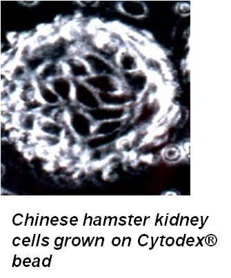
All these issues are resolved when microfluidic manipulation is used to handle live cells grown on beads such as Cytodex® or Cytopor®.


Reaching the present state of technology took many years, yet an important milestone was reached at an early stage, when it was shown that cells, cultured on carrier beads, can be reliably transported within the SI system, and monitored by fluorescence microscopy (Hodder 1999, 2000). Although the most recent developments of fluorescence, absorbance, and electrochemical probing of cellular responses in the microSI-LOV system (Lähdesmäki 2009) makes the use of fluorescence microscopy obsolete, the following sections include our previous work with microscopy, since they demonstrate the principles and reveal novel observations on the kinetics of cellular responses that could not be obtained in any other way. Also, It will be easy to adapt the cellular assays, developed for fluorescence microscopy, into less cumbersome and more versatile microSI-LOV systems.
Ruzicka, J.; Lindberg W.;. Anal. Chem. 1992, 64, 537A.